Independent Provider of Microscopy and Imaging Solutions in the Nordics
Visi4
Material Microscopy for research, inspection, measurement and analysis
Visi4 can offer Digital Inspection Systems and Optical Measurement Systems for a wide range of applications. Our main purpose is to provide a custom-tailored solution to suit the individual requirements.
Visi4 selects camera, stands or microscope, optics, illumination and software - creating a complete material imaging system - to fulfil your specifications of demands also whether you need a manual or an automated imaging system.
Visi4 has experience with imaging applications i.e raw materials, powder, biomaterials, polymers, metals, geomaterials and electronics in QA/QC in industry and manufacturing as well as material research.
CONTACT US
Info@Visi4.com
Tel: +45 61666619
Peter Thomsen
Innovation &
Microscopy expert.
Stengårds Alle 31A
2800 Lyngby
Denmark
PROFILE
Visi4 commercialize advanced imaging solutions.
These solutions are proven in Visi4's development of vascular implants.
FOLLOW Visi4
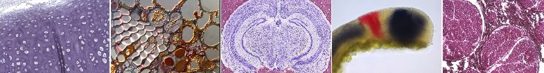
Histology and Fluorescence Microscopy
The histology or histopathology is the study of microscopic anatomy of cells, tissues, diseased tissue or cancer. It is performed by examining the sample under a light microscope. The ability to visualize or differentially identify microscopic structures is the main challenge for the Visitron HistoScope Imaging System. To get the best resolution and true colour information for the correct analysis and diagnosis we use state of the art colour CCD cameras with three different technologies. Colour mosaic CCD, Pixel shifting CCD technique and liquid crystal three shot sequential RGB channel acquisition. The easy to use VisiView imaging software, developed by Visitron Systems GmbH controls the camera acquisition, colour correction, overlay and analysis of the images. In addition motorized microscopes are also supported e.g. for screening of cells or tissue.
The majority of fluorescence microscopes, especially those used in the life sciences, are of the epifluorescence design. Light of the excitation wavelength is focused on the specimen through the objective lens. The fluorescence emitted by the specimen is focused to the detector by the same objective that is used for the excitation which for greater resolution will need objective lens with higher numerical aperture. Since most of the excitation light is transmitted through the specimen, only reflected excitatory light reaches the objective together with the emitted light and the epifluorescence method therefore gives a high signal-to-noise ratio. An additional wavelength specific filter between the objective and the detector can filter out the remaining excitation light from fluorescent light. Visi4 provides high effiency filter sets and intelligent microscope illumination systems which match you applications and fits to your imaging system.
Visi4 offers fiber coupled HXP/XCite - DG4/5 - High Speed Polychromatic Illumination - CoolLed.